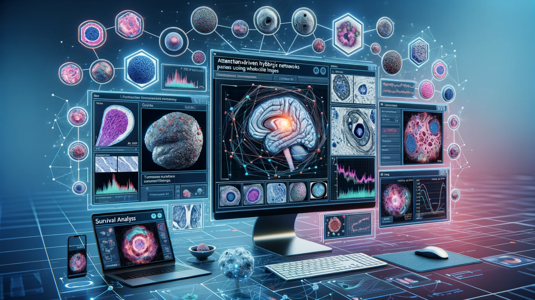
An Attention-Driven Hybrid Network for Survival Analysis of Tumorigenesis Patients Using Whole Slide Images December 27, 2023 by Vision @Seecs AI For Medical Image Analytics
Attention-aware Feature Fusion based Nuclei Instance Segmentation and Type Classification Using Histology Images June 8, 2023 by Vision @Seecs AI For Medical Image Analytics
AI-Powered Cephalometric Landmark Detection for Orthodontic Diagnosis: Framework, Dataset, and Detection Challenge June 8, 2023 by Vision @Seecs AI For Medical Image Analytics
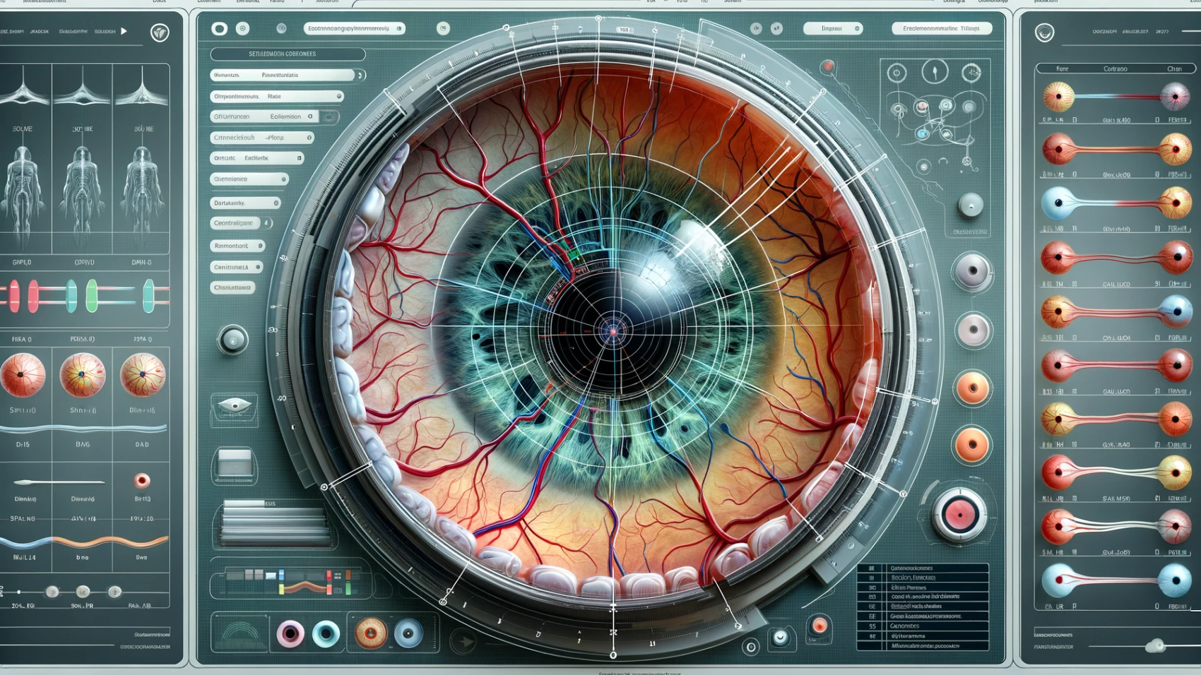
Detection & segmentation of anatomical structures in retinal images March 1, 2022 by AI For Medical Image Analytics
Cellular community detection for tissue phenotyping in digital histology images March 1, 2022 by AI For Medical Image Analytics
Segmentation of micro-vessels and nerves in digital histology images February 28, 2022 by AI For Medical Image Analytics
Computational Pathology for improved cancer management and better human sustainability February 27, 2022 by AI For Medical Image Analytics